He Saw Through His Wife’s Hand and the World Was Never the Same: The Accidental Discovery That Launched Modern Medicine
On a November evening in 1895, a German physicist working alone in a darkened laboratory stumbled upon something that shouldn’t have been possible. Within weeks, medicine would never be the same. Within years, men would die for it.
Six Weeks of Obsessive Secrecy
Wilhelm Conrad Röntgen was not a man prone to drama. By November 1895, the fifty-year-old professor of physics at the University of Würzburg had built a respectable but unremarkable career. He published careful papers, taught conscientious students, and worked in the methodical tradition of German natural science. He was not the sort of man who changed the world.
And then, on the evening of November 8, 1895, he did exactly that.
Röntgen was experimenting with a Crookes tube — a glass vacuum tube through which electrical current could be discharged — a standard piece of apparatus for physicists studying cathode rays in the 1890s. His laboratory was darkened. The tube was wrapped in black cardboard to block any light it might emit. And yet, roughly a meter away, a small fluorescent screen coated with barium platinocyanide began to glow.
It should not have glowed. Nothing in Röntgen’s understanding of physics could explain it. Cathode rays, the phenomenon he was studying, could travel only a few centimeters through air. Whatever was crossing the room to illuminate that screen was something else entirely.
What followed was one of the most extraordinary episodes of private scientific obsession in recorded history. Röntgen told no one — not his colleagues, not his department, not his wife. He moved a cot into the laboratory and spent approximately six weeks barely sleeping and barely eating, methodically testing every property of this invisible radiation he had stumbled upon. He passed it through paper, through wood, through books. He found it could expose photographic plates. He discovered it traveled in straight lines and could not be deflected by magnets, distinguishing it fundamentally from the cathode rays he’d been studying.
He named it the X-ray — X for the mathematical unknown — a deliberately provisional name that acknowledged he didn’t yet understand what he had found. In most languages, the name stuck. In German, it became Röntgenstrahlen, Röntgen rays, giving the discoverer a nominal immortality that English speakers largely denied him.
“I Have Seen My Death”
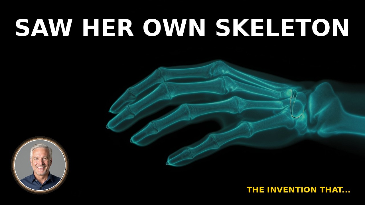
When Röntgen finally felt confident enough to demonstrate his discovery, he turned to the person he trusted most. On December 22, 1895, he asked his wife, Anna Bertha Ludwig, to place her hand flat on a photographic plate. He exposed it for fifteen minutes. When the image developed, it showed the bones of her hand with crystalline clarity, her wedding ring sitting dense and opaque against the ghostly architecture of her skeleton.
Anna Bertha Röntgen reportedly said she had seen her own death.
The remark has echoed through history partly because it was so prophetic — not for her specifically, but for the story that would follow. She was confronting something humans had never confronted before: themselves, rendered transparent. The Victorian certainty that the body was a private, opaque vessel had been dissolved in a single photographic exposure.
Röntgen submitted his paper, On a New Kind of Ray, to the Würzburg Physical-Medical Society on December 28, 1895 — barely seven weeks after his initial discovery. He included prints of the hand radiograph. When the paper reached the international scientific community in early January 1896, the response was immediate and global. Within a month, X-ray laboratories were being established across Europe and North America. Within a year, military surgeons were using the technology to locate bullets in wounded soldiers.
The speed of adoption was almost without precedent in the history of medicine.
The Physics of Seeing Through Flesh
Understanding why X-rays work requires a brief detour into the electromagnetic spectrum — the continuous range of radiation that includes visible light, radio waves, and gamma rays, organized by wavelength and energy.
Visible light, the radiation our eyes evolved to detect, has wavelengths between roughly 380 and 700 nanometers. X-rays have wavelengths between 0.01 and 10 nanometers, far shorter and correspondingly far more energetic. This higher energy is what gives them their penetrating power.
When X-rays pass through human tissue, different materials absorb them at different rates. Soft tissue — muscle, fat, organs — is largely transparent to X-rays, allowing them to pass through and expose the photographic film behind. Dense materials — bone, metal, heavily calcified tissue — absorb significantly more radiation and appear opaque or white in the resulting image. The gradations between these extremes produce the ghostly topography of the radiograph.
What Röntgen couldn’t have known in 1895 was that this penetrating power came with a cost. X-rays are ionizing radiation — they carry enough energy to strip electrons from atoms, breaking chemical bonds and damaging biological tissue at the cellular level. In small doses, the damage is negligible and the diagnostic benefit vastly outweighs the risk. In the chronic, unprotected exposures that characterized the first decade of X-ray experimentation, the consequences could be catastrophic.
The Men Who Died for the Discovery
If Röntgen’s story is one of private triumph, the story of early X-ray workers is one of public tragedy.
Thomas Edison, working from his laboratory in West Orange, New Jersey, launched an aggressive X-ray research program in early 1896. He assigned a young glassblower named Clarence Dally to the project, tasking him with developing improved fluorescent materials for X-ray screens. Dally was talented and diligent. He was also, in the course of his work, exposing his hands to X-rays for hours at a time, day after day, without any protection whatsoever.
The damage was cumulative and horrific. Dally developed radiation burns that progressed to ulceration, then to cancer. His left hand was amputated. Then his right. Then his arms. He died in 1904 at the age of thirty-nine, becoming the first recorded death attributed to radiation exposure in the United States. Edison, shaken by what he witnessed, abandoned his X-ray research entirely and reportedly refused to discuss it for the rest of his life.
Dally was not alone. Across Europe and America, X-ray pioneers developed what they called “X-ray dermatitis” — a term that covered everything from persistent skin redness to gangrenous destruction of tissue. Dozens of early radiologists lost fingers, hands, and arms. Many died of radiation-induced cancers. A monument erected in Hamburg in 1936 lists 169 names — researchers and physicians from fifteen countries who died as martyrs to the new science.
The terrible irony is that many of these casualties occurred not through ignorance of the danger, but despite increasingly clear warnings. As early as 1896, researchers were noting skin damage from X-ray exposure. The science simply moved faster than the culture of caution could follow.
The Nobel Prize and the Question of a Patent
In 1901, Wilhelm Röntgen received the very first Nobel Prize in Physics, the committee citing his discovery of “the remarkable rays subsequently named after him.” The award formalized what scientists had recognized almost immediately: that the discovery of X-rays was among the most consequential in the history of physics.
Röntgen’s response to his discovery’s extraordinary commercial and scientific value is worth sitting with. He refused to patent X-rays or the equipment used to produce them. He believed, in a manner deeply characteristic of a certain tradition of European academic science, that scientific discoveries belonged to humanity rather than to individuals or corporations. He never sought to profit from X-rays. He donated his Nobel Prize money to the University of Würzburg.
This decision stands in sharp contrast to the competitive, patent-driven approach that characterized much American industrial research of the same period — the Edison model, in which discovery and profit were understood as inseparable. The contrast raises genuinely difficult questions about how scientific progress actually happens.
The argument for patents is intuitive: profit incentives drive investment, investment accelerates development, and competition breeds efficiency. Had Röntgen controlled X-ray technology, the argument goes, a well-funded corporate program might have advanced the science faster than the diffuse, underfunded efforts of university researchers worldwide.
The argument against is equally compelling. X-rays spread across the globe in months precisely because there were no barriers to adoption. Military surgeons in the Sudan and Greece were using the technology within a year of its discovery. Medical schools could acquire and experiment with equipment without licensing fees or legal complications. The speed of diffusion was itself a form of accelerated development — thousands of researchers worldwide, rather than a single well-funded laboratory, were simultaneously discovering applications.
What the historical record suggests, though cannot prove, is that open science won the 1890s. The pace of X-ray development in that first decade — from discovery to clinical adoption to the first dedicated radiology departments — is nearly unmatched in medical history.
A Pattern Older Than X-Rays
Röntgen’s discovery belongs to a surprisingly large category of civilization-altering accidents. Penicillin emerged from a contaminated Petri dish in 1928. Teflon was discovered when a refrigerant experiment went wrong in 1938. Radioactivity itself — the broader phenomenon of which X-rays are one expression — was accidentally discovered by Henri Becquerel in 1896, when a cloudy Parisian week prevented him from conducting the experiment he actually intended.
What these accidents share is not randomness but what the French scientist Louis Pasteur called “the prepared mind” — the researcher who, encountering something unexpected, has the knowledge, curiosity, and freedom to follow it rather than dismiss it. Röntgen noticed the fluorescent screen because he was the kind of scientist who noticed things. He spent six weeks investigating it because he worked in an institution that gave him the freedom to do so.
Modern medical imaging — the CT scanner, the MRI, the PET scan, the fluoroscope — descends in a direct line from that November evening in Würzburg. The bones of Anna Bertha Röntgen’s hand, captured in a fifteen-minute exposure in December 1895, were the first image in a visual vocabulary that physicians now use billions of times each year.
She said she had seen her own death. What she had actually seen was medicine learning to see itself.
This article accompanies a documentary episode exploring the discovery of X-rays, the physics of electromagnetic radiation, and the human cost of Victorian scientific experimentation. The history of Wilhelm Röntgen, Clarence Dally, and the early pioneers of radiology draws on primary sources including Röntgen’s 1895 paper, contemporary accounts in Nature and Science*, and the historical archives of the German Röntgen Museum in Remscheid.*